A high-resolution nanoscale window to the live biological world
December 27, 2012

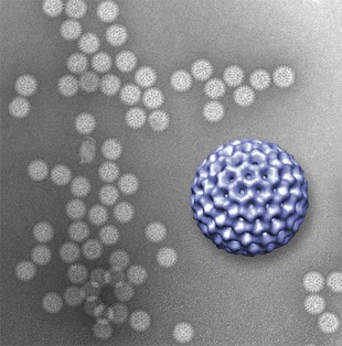
A novel microfluidics platform allowed viewing of structural details of rotavirus double-layered particles; the 3-D graphic of the virus, in purple, was reconstructed from data gathered by the new technique and is ~80 nm. in diameter (credit: Virginia Tech)
Investigators at the Virginia Tech Carilion Research Institute haveinvented a way to directly image biological structures at nanometer-resolution in their natural habitats (a liquid environment).
The technique is a major advancement toward the ultimate goal of imaging biological processes in action at the atomic level.
The technique uses two silicon-nitride microchips with windows etched in their centers and pressing them together until only a 150-nanometer space between them remains.
The researchers then fill this pocket with a liquid resembling the natural environment of the biological structure to be imaged, creating a microfluidic chamber.
Then, because the movement of free-floating structures yield images with poor resolution, the researchers coat the microchip’s interior surface with a layer of natural biological tethers, such as antibodies, which naturally grab onto a virus and hold it in place./.../
No comments:
Post a Comment