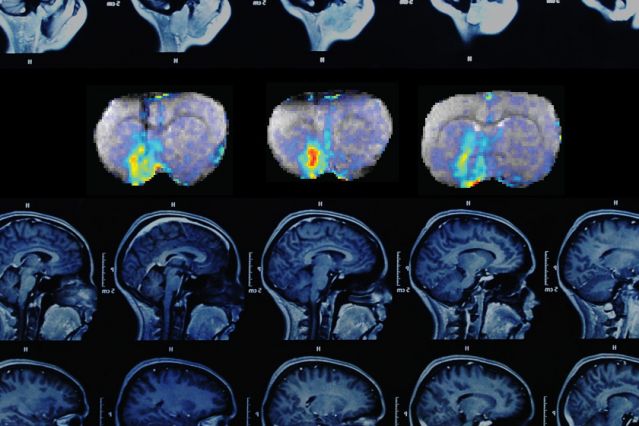
A series of three MRI images (top row) shows how dopamine concentrations change over time in the brain’s ventral striatum. (Credit: photocollage by Christine Daniloff/MIT, with images courtesy of the researchers)
MIT researchers have developed a technique that allows them to precisely track neural communication in the brain over time, using magnetic resonance imaging (MRI) along with a specialized molecular MRI contrast agent.
This is the first time anyone has been able to map neural signals with high precision over large brain regions in living animals, offering a new window on brain function, says Alan Jasanoff, an MIT associate professor of biological engineering and an associate member of MIT’s McGovern Institute for Brain Research.
His team used this molecular imaging approach, described in the May 1 online edition of Science, to study the neurotransmitter dopamine in a region called the ventral striatum, which is involved in motivation, reward, and reinforcement of behavior. In future studies, Jasanoff plans to combine dopamine imaging with functional MRI techniques that measure overall brain activity to gain a better understanding of how dopamine levels influence neural circuitry.
“We want to be able to relate dopamine signaling to other neural processes that are going on,” Jasanoff says. “We can look at different types of stimuli and try to understand what dopamine is doing in different brain regions and relate it to other measures of brain function.”/.../
No comments:
Post a Comment