A 3D window into living cells, no dye required
January 27, 2014

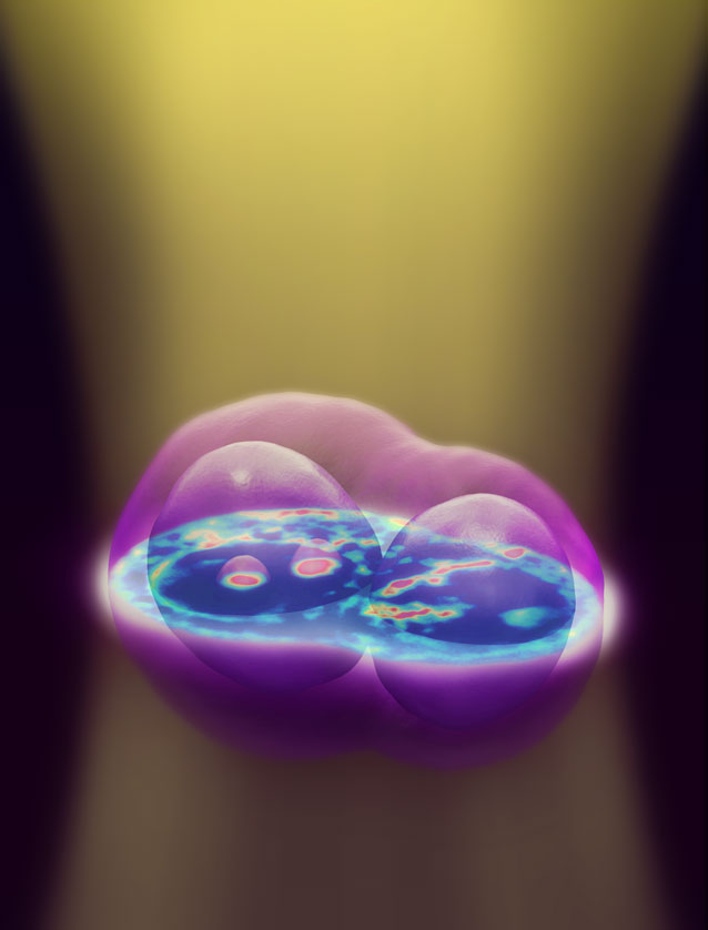
A new 3D imaging technique for live cells uses a conventional microscope to capture image slices throughout the depth of the cell, then computationally renders them into one three-dimensional image. The technique uses no dyes or chemicals, allowing researchers to observe cells in their natural state. (Credit: University of Illinois)
University of Illinois researchers have developed a new imaging technique that needs no dyes or other chemicals, yet renders high-resolution, three-dimensional, quantitative imagery of cells and their internal structures using conventional microscopes and white light.
Called white-light diffraction tomography (WDT), the imaging technique opens a window into the life of a cell without disturbing it and could allow cellular biologists unprecedented insight into cellular processes, drug effects and stem cell differentiation.
The team, led by electrical and computer engineeringand bioengineering professor Gabriel Popescu, published their results in the journal Nature Photonics./.../
No comments:
Post a Comment